Your shoulder pain, rounded posture, and restricted breathing may share one source — and stretching it isn’t enough
There’s a particular kind of frustration that comes from doing everything right and still hurting. You’ve stretched your chest. You’ve foam-rolled your upper back. You’ve done the rotator cuff exercises your physical therapist gave you. The shoulder pain comes back. The stiffness between your shoulder blades doesn’t move. And somewhere along the way, you noticed that taking a deep breath feels harder than it should — like something is holding your ribs in place.
These feel like separate problems. They’re usually treated as separate problems. But there’s a reasonable chance they share a single source: a small, triangle-shaped muscle buried under your pectoralis major that most people have never heard of and almost no one thinks to look at.
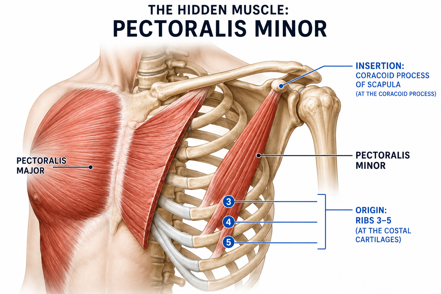
The pectoralis minor connects your ribs to your shoulder blade. It sits directly on top of the nerves and blood vessels that supply your entire arm. And when it gets tight — which it does, easily and often — it sets off a chain of mechanical consequences that scatter pain, restriction, and numbness across your upper body in ways that get chased individually for months or years without resolution.
Understanding this muscle — what it does, why it tightens, what happens when it does, and why the standard advice doesn’t fix it — is the difference between treating symptoms and actually solving the problem.
What the Pectoralis Minor Does
The pectoralis minor is a flat, triangular muscle that originates from the front surfaces of your third, fourth, and fifth ribs, near where bone meets cartilage. Its fibers angle upward and outward to attach to a small hook-shaped piece of bone on the front of your shoulder blade called the coracoid process. This arrangement means the muscle does one primary thing: it pulls your shoulder blade forward, downward, and tight against your ribcage.
That sounds simple, but the implications are not. The scapula — your shoulder blade — is a floating bone. It’s not locked to your skeleton by a joint the way your femur is locked to your pelvis. It moves across the back of your ribcage on a layer of muscle, and its position determines everything about how your shoulder functions. The pec minor is one of the muscles that controls that position. When it contracts, it anchors the scapula. When it stays contracted, it anchors the scapula in the wrong place.
There’s a second role that most anatomy articles mention in passing and then move on from, even though it matters enormously: the pectoralis minor is an accessory muscle of respiration. When your ribs are held stable from below, the muscle pulls the shoulder blade down. But when the shoulder blade is held stable from above, the same contraction lifts the ribs — expanding the chest cavity, pulling air in. This is your emergency breathing system. When you need to gasp — during heavy exertion, respiratory distress, or panic — the pec minor kicks in and lifts your ribcage to get more air.
The problem is that many people have turned their emergency breathing system into their primary one.
How the Pectoralis Minor Gets Tight — And Why “Bad Posture” Is Not the Full Answer
Every article on this muscle mentions posture. Sitting at a desk rounds your shoulders forward, shortening the pec minor over time, and eventually the muscle adapts to the shortened position. This is true. It’s also incomplete.
There are at least three distinct pathways to a tight pectoralis minor, and they require different solutions. Lumping them all under “posture” is why the generic advice doesn’t stick.
The first pathway is anterior dominance. The person pushes more than they pull — in the gym, in their daily movement patterns, in how they carry loads. The pec minor gets disproportionately strong relative to its antagonists on the back of the shoulder blade. It wins the tug-of-war and pulls the scapula forward at rest, not because it’s injured or dysfunctional, but because nothing on the other side is strong enough to pull back. This is common in people who bench press frequently, do a lot of push-ups, or carry heavy objects in front of their body.
The second pathway is compensation. The muscles that are supposed to stabilize the scapula — primarily the lower trapezius and the serratus anterior — are weak. The body doesn’t tolerate an unstable scapula, so it recruits the next available muscle to lock it down. That muscle is the pec minor. It wasn’t designed for sustained stabilization work, but the body doesn’t care about design — it cares about getting through the day. The pec minor tightens because it’s doing someone else’s job, and it will stay tight for as long as that job goes unfilled.
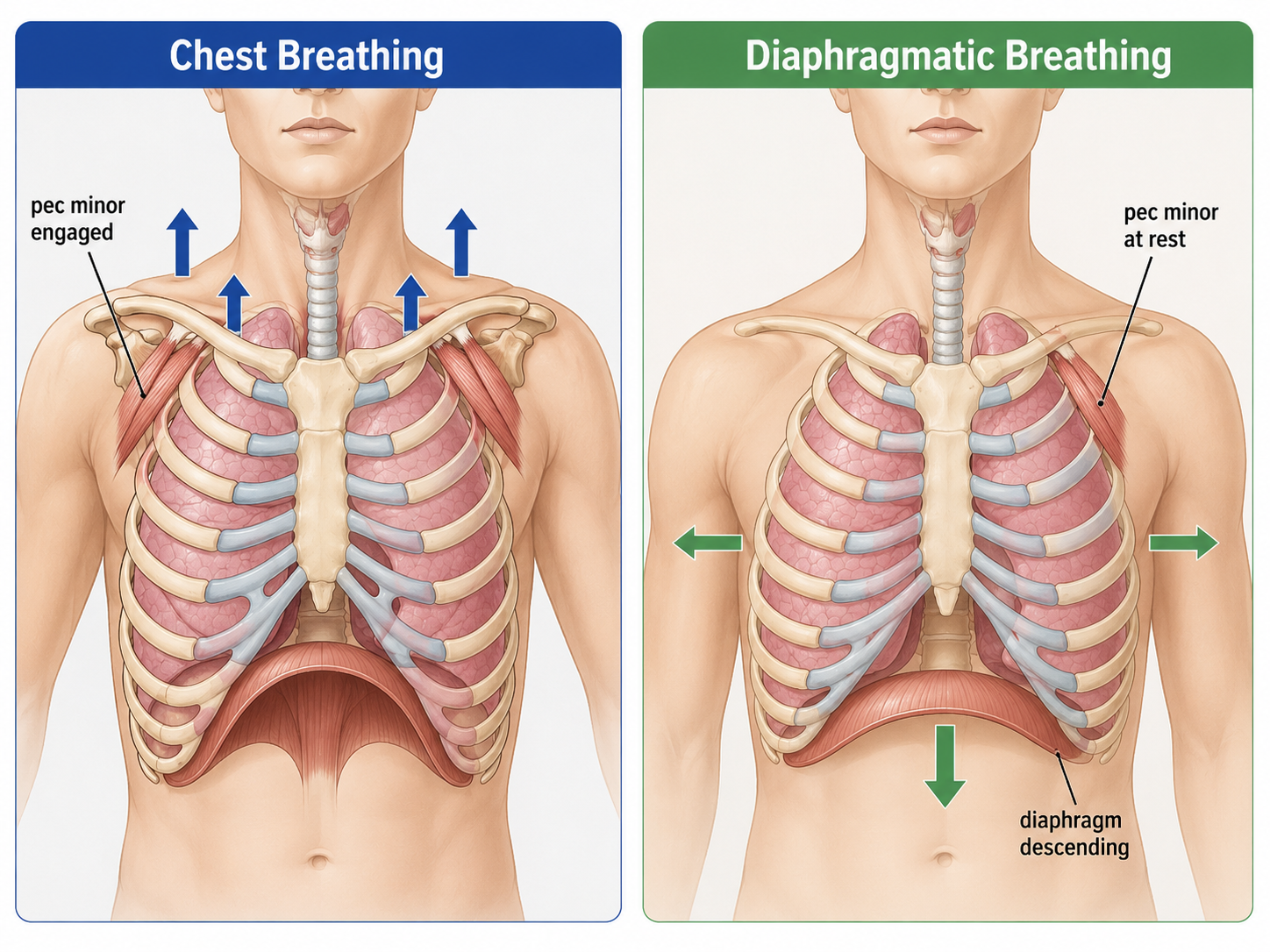
The third pathway is the one most articles skip almost entirely: breathing pattern dysfunction. When you breathe primarily with your chest — ribs lifting up instead of expanding out, shoulders rising on each inhale, belly staying flat — the pec minor is contracting on every single breath cycle. At a normal resting breathing rate, that’s roughly 15,000 contractions per day in a role the muscle was built for occasional use. Chronic shallow breathing is not just a stress symptom; it’s a mechanical load on the pec minor that makes the muscle progressively tighter and more painful regardless of your posture or your gym program.
This is where the relationship between anxiety and chest tightness becomes something more specific than “stress causes tension.” Anxiety changes breathing patterns. Changed breathing patterns recruit the pec minor. The pec minor tightens. The tight muscle restricts rib expansion. Restricted rib expansion makes deep breathing harder. Harder breathing reinforces the anxiety. It’s a loop with a measurable muscular mechanism at its center.
What a Tight Pectoralis Minor Actually Does to Your Body
When the pec minor shortens, the consequences don’t stay local. They cascade.
The first thing that changes is scapular position. The shoulder blade tips forward — its bottom edge pokes out from the back (sometimes called scapular winging), and the socket of the shoulder joint (the glenoid cavity) loses its normal upward orientation. You can sometimes see this asymmetry by comparing how far the front of each shoulder sits from the chest.
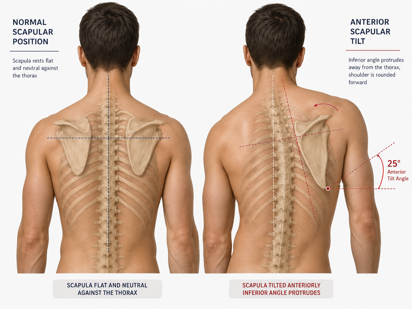
This tilt changes shoulder mechanics. When you raise your arm overhead, the scapula needs to rotate upward to keep the shoulder joint aligned. A forward-tilted scapula can’t complete that rotation. The arm runs out of room, and the rotator cuff tendons get pinched between the top of the arm bone and the bony shelf above the socket — the acromion. This is subacromial impingement, and it’s one of the most common shoulder diagnoses in the world. It often gets treated as a rotator cuff problem. If the underlying cause is a pec minor that won’t let the scapula rotate, the rotator cuff exercises will provide temporary relief at best.
The scapular tilt also changes what happens further up the chain. When the shoulder blades pull forward, the upper thoracic spine rounds to accommodate them. The increased curvature of the upper back forces the neck into extension to keep the eyes level — the head juts forward, the suboccipital muscles at the base of the skull tighten, and the upper trapezius works overtime to hold everything in position. The result is upper back stiffness, neck pain, and in some cases, tension headaches. This pattern — tight pec minor, rounded upper back, forward head, tight upper traps — is often described as upper crossed syndrome. The pec minor is not the only contributor, but it’s frequently the initiating one.
The most serious consequence involves the structures that run directly beneath the muscle. The three cords of the brachial plexus — the nerve bundle that supplies your entire arm — and the axillary artery and vein all pass through a space between the pec minor and the ribcage. When the muscle shortens, that space narrows. The nerves and vessels get compressed.
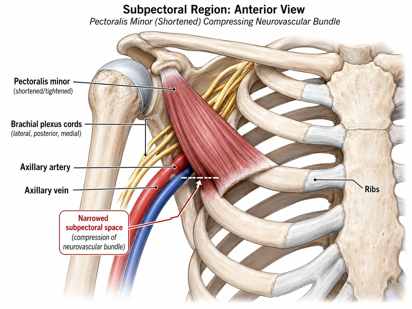
This compression produces symptoms that are easily mistaken for other conditions: numbness and tingling down the arm and into the hand, particularly the ring and little fingers; a feeling of heaviness or swelling in the arm; coldness or color changes in the hand; and pain in the shoulder, chest, or armpit that can mimic cardiac symptoms. This is pectoralis minor syndrome, a subset of thoracic outlet syndrome that is frequently misdiagnosed or overlooked entirely. Patients often get worked up for cervical radiculopathy, carpal tunnel syndrome, or rotator cuff tears before anyone looks at the pec minor.
Why Stretching Alone Doesn’t Fix It
This is the central point that most advice on this muscle misses, and it’s the reason people stretch their pec minor for weeks or months and the tightness keeps returning.
If the muscle is tight because it’s compensating for weak scapular stabilizers, stretching it removes the only thing currently holding the scapula in place. The body registers the instability and re-tightens the muscle to protect itself. You’ve loosened a compensation without fixing the deficit it was compensating for. The tightness returns, sometimes within hours.
If the muscle is tight because of a breathing pattern issue, stretching does nothing about the 15,000 daily contractions that are maintaining the problem. You stretch for thirty seconds; the muscle contracts on the next breath and the one after that and the one after that.
If the muscle is tight because of anterior dominance, stretching temporarily lengthens it, but the strength imbalance that pulled it short in the first place hasn’t changed. It drifts back to its shortened position under the pull of the stronger front-side muscles.
Stretching is not wrong. It’s incomplete. It addresses the state of the muscle — its current length — without addressing the reason it arrived at that length. The fix has to go deeper.
What Actually Works
The solution matches the cause, and in most cases, all three layers need attention.
Breathing retraining is the intervention that gets left out of almost every pec minor article, despite being the one that changes the loading pattern most dramatically. The goal is straightforward: shift the primary work of breathing from the chest to the diaphragm. When the diaphragm contracts, the ribs expand laterally and the belly moves outward. The pec minor stays quiet. The cue that matters most is lateral rib expansion — the ribs moving out to the sides, not upward — which is the signature of diaphragmatic breathing and the direct opposite of the pattern that overloads the pec minor. This is not a relaxation technique bolted onto a rehab program. It’s a mechanical unloading of the muscle that stretching cannot accomplish.
Scapular stabilizer strengthening fills the gap the pec minor has been compensating for. The serratus anterior is the priority — it protracts the scapula and holds it against the ribcage, the same job the pec minor has been doing under protest. The serratus punch and the push-up plus (a push-up with an extra protraction at the top, emphasizing the concentric phase) are both highly selective for serratus anterior activation with minimal pec minor involvement. Lower trapezius training — prone Y-raises, wall slides with posterior tilt — restores the upward rotation force that the tight pec minor has been blocking. Rhomboid work supports both. The goal is to give the scapula real stabilizers so the pec minor can stand down.
Pec minor release and stretch — now, with the cause addressed, lengthening the muscle actually holds. Self-palpation is possible but requires some feel: find the coracoid process (the bony point just below the outer end of your collarbone, in the front of your shoulder), then press just below and slightly inward. The pec minor lies beneath the pec major here, and if it’s tight, you’ll feel a firm, tender band when you push through. A doorway stretch with the elbow above shoulder height (which biases the stretch toward the pec minor rather than the pec major) is the classic lengthening approach. Manual therapy — direct pressure, soft tissue mobilization — can accelerate release for muscles that have been in sustained contraction.
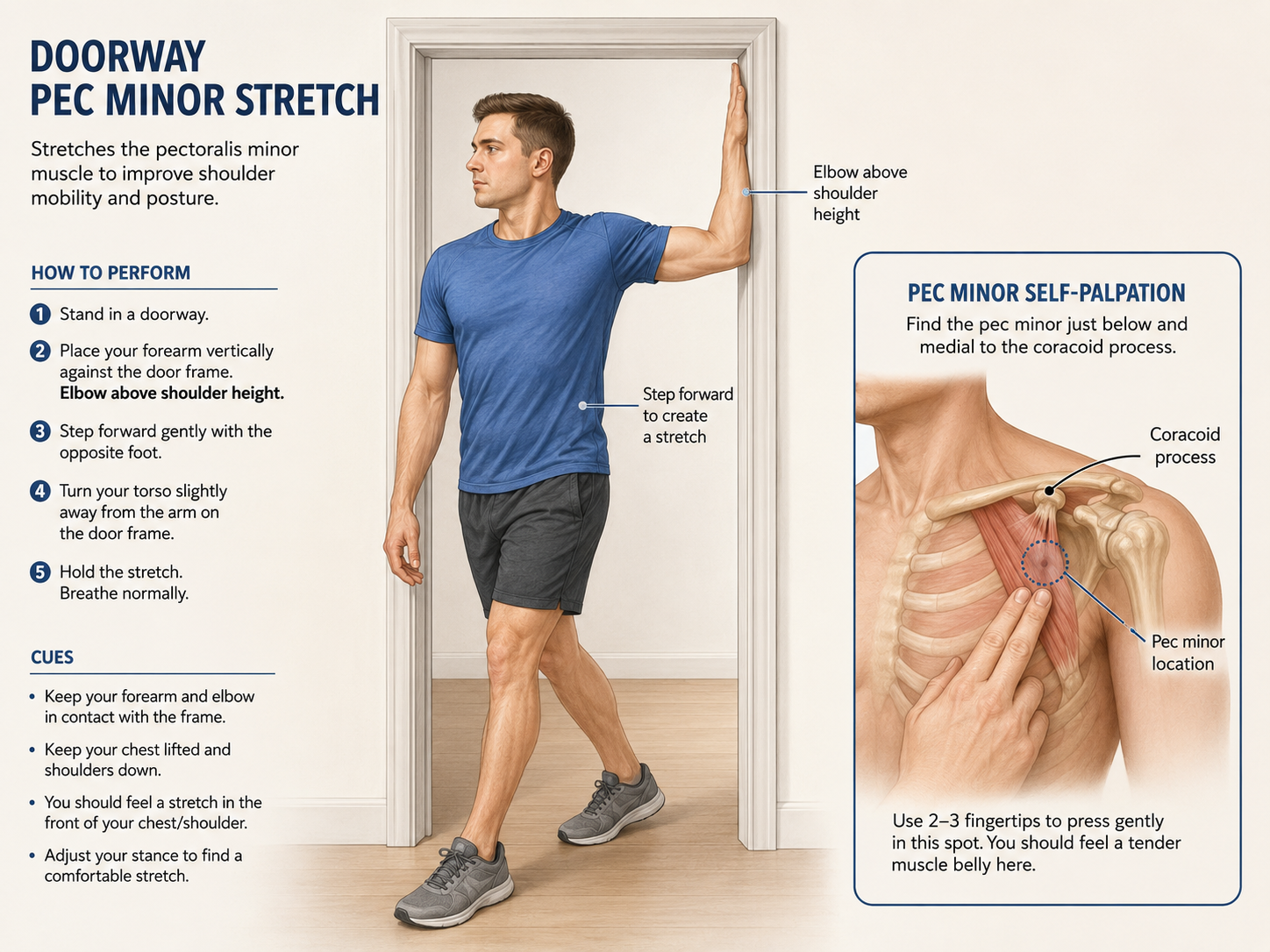
Movement pattern correction integrates the work. Overhead reaching, pulling, and pushing with a scapula that now has the freedom and the muscular support to track correctly. Without this step, the isolated gains from breathing work, strengthening, and stretching don’t transfer into the movements that caused the problem.
When to See a Professional
If you have numbness, tingling, or loss of sensation in your arm or hand; if you notice swelling, coldness, or color changes in one arm; if chest pain is involved in any way — these are not self-treatment situations. Pectoralis minor syndrome involves compression of nerves and blood vessels that requires clinical assessment to diagnose properly. A diagnostic ultrasound-guided muscle block — where a local anesthetic is injected into the pec minor under imaging — can confirm whether the muscle is the source of compression. Imaging can rule out structural causes. And in rare cases where conservative treatment fails, surgical release of the pec minor tendon from the coracoid process has shown consistent results.
The important thing is knowing these options exist, because many clinicians don’t have pectoralis minor syndrome on their differential diagnosis list. If you’ve been worked up for rotator cuff pathology, cervical disc issues, or carpal tunnel and nothing has explained your symptoms, it’s worth asking whether anyone has looked at the pec minor.
The Muscle That Connects Everything
The pectoralis minor doesn’t show up in mirror checks. It doesn’t appear on most gym programs. It doesn’t come up in casual conversation about shoulder pain. But it sits at the exact intersection of three systems that govern how your upper body feels and functions: your breathing mechanics, your scapular position, and your neurovascular supply to the arm.
When it works properly, you never think about it. When it doesn’t, it produces a pattern of symptoms — shoulder pain, upper back stiffness, restricted breathing, tingling hands — that look unrelated and get treated separately, often for a long time, often without resolution.
The fix isn’t complicated. But it does require understanding the muscle as part of a connected system rather than an isolated structure to stretch. Address why it’s tight — the breathing pattern, the scapular weakness, the anterior dominance — and the tightness resolves. Address only the tightness, and you’ll be stretching it again next week.